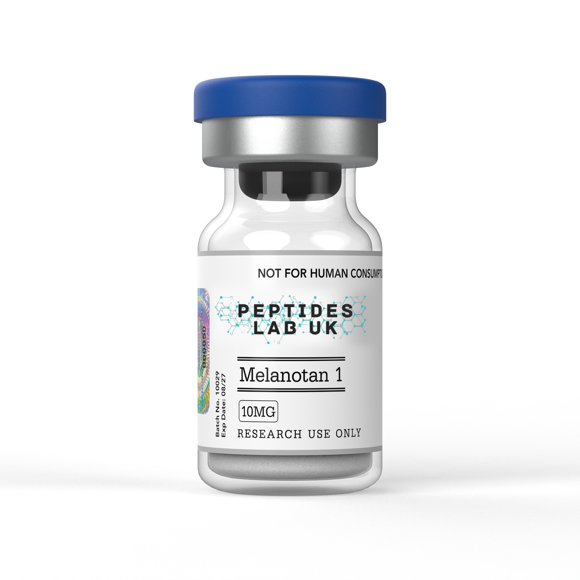
Melanotan 1, chemically designated [Nle4, D-Phe7]-alpha-MSH (NDP-MSH) and known by its international nonproprietary name afamelanotide, is a synthetic linear heptapeptide analogue of endogenous alpha-melanocyte-stimulating hormone. Two targeted amino acid substitutions, replacement of methionine at position 4 with norleucine and phenylalanine at position 7 with D-phenylalanine, confer substantially greater resistance to enzymatic degradation and higher potency than the native hormone. Melanotan 1 shows preferential selectivity for the melanocortin-1 receptor (MC1R) expressed on epidermal melanocytes, driving intracellular cAMP-dependent signalling that activates the eumelanin synthesis pathway. Within laboratory research, melanotan 1 is used to study MC1R receptor pharmacology, melanogenesis signalling cascades, and photoprotective mechanisms in melanocyte cell and tissue models.
What Is Melanotan 1?
Melanotan 1 was developed through structural modification of native alpha-MSH following the identification of the minimal amino acid sequence, His6-Phe-Arg-Trp9, required for melanocortin receptor biological activity. Building on this minimal active core, researchers introduced two specific substitutions to the full 13-amino-acid alpha-MSH sequence: methionine at position 4 was replaced with norleucine, preventing oxidation of the native methionine residue and thereby improving chemical stability, while phenylalanine at position 7 was replaced with D-phenylalanine, a stereochemical modification that substantially increases resistance to enzymatic proteolysis relative to the all-L-amino-acid native hormone. Together these changes produced NDP-MSH, a peptide with markedly higher potency and metabolic stability than native alpha-MSH.
This enhanced stability translates directly into an extended functional half-life relative to native alpha-MSH, which is rapidly degraded in circulation. The specific combination of the D-amino acid substitution at position 7, which confers resistance to enzymatic cleavage, and the norleucine substitution at position 4, which prevents oxidative degradation, together account for melanotan 1’s substantially prolonged duration of receptor engagement compared with the native hormone, a structural principle shared with numerous other stabilised peptide hormone analogues developed using similar D-amino-acid substitution strategies.
Binding affinity profiling across the melanocortin receptor family (MC1R through MC5R) has shown that melanotan 1 retains activity across multiple receptor subtypes but demonstrates preferential engagement of MC1R, the subtype expressed predominantly on epidermal melanocytes and responsible for regulating pigmentation. This receptor selectivity profile has been further clarified through structural biology research using cryo-electron microscopy to resolve the MC1R-Gs protein complex bound to several ligands, including endogenous alpha-MSH, afamelanotide itself, and other synthetic melanocortin agonists, providing atomic-level structural detail relevant to understanding the basis for melanotan 1’s receptor engagement and informing broader efforts to resolve receptor-selectivity challenges across melanocortin-targeted drug discovery research.
Structural stability considerations extend beyond simple degradation resistance to encompass the peptide’s overall research handling profile. Because melanotan 1 retains the fundamental heptapeptide-derived pharmacophore responsible for melanocortin receptor engagement while incorporating targeted stability-enhancing substitutions, it has become a valuable structural reference point in melanocortin receptor structure-activity research, distinguishing it from both the native, rapidly degraded alpha-MSH hormone and from more extensively modified synthetic analogues such as Melanotan 2, which incorporates a cyclic lactam bridge structure and shows a broader, less MC1R-selective receptor engagement profile.
Mechanism of Action
Melanotan 1’s principal mechanism begins with selective binding to MC1R, a class A G-protein-coupled receptor expressed on the surface of epidermal melanocytes. Upon ligand binding, MC1R couples to the heterotrimeric Gs protein, and receptor activation triggers dissociation of the Gαs subunit, which subsequently activates adenylyl cyclase. This enzymatic activation catalyses the conversion of ATP to cyclic adenosine monophosphate (cAMP), establishing cAMP as the crucial second messenger governing downstream melanogenic signalling following melanotan 1-MC1R engagement.
Elevated intracellular cAMP activates protein kinase A (PKA), which subsequently phosphorylates the cAMP response element-binding protein (CREB). Phosphorylated CREB drives transcriptional activation of the microphthalmia-associated transcription factor (MITF) gene, positioning MITF as the principal downstream transcriptional regulator of the entire melanogenic gene programme triggered by MC1R activation. MITF is widely recognised in the pigmentation research literature as the master transcriptional regulator of melanin biosynthesis, and its activation represents the central node through which melanotan 1’s receptor-level signalling is translated into the cellular machinery required for melanin production.
MITF activation initiates transcription of numerous genes encoding melanogenesis-associated proteins, most centrally tyrosinase (TYR), alongside tyrosinase-related protein 1 (TYRP1) and dopachrome tautomerase (DCT). Tyrosinase occupies a particularly critical position within this enzymatic cascade, since it catalyses the oxidation of tyrosine to dopaquinone, the first and rate-limiting step of the entire melanogenesis pathway. TYRP1 and DCT participate in subsequent steps of melanin synthesis and additionally play regulatory roles in organising the broader melanogenic apparatus within the melanocyte. Research has confirmed that melanotan 1 upregulates tyrosinase gene expression and directly increases tyrosinase enzymatic activity in treated melanocytes, in addition to its upstream effects on MITF transcriptional activation, indicating that melanotan 1’s melanogenic effects operate through convergent action at multiple points in this enzymatic cascade rather than through MITF transcriptional upregulation alone.
The end product of this signalling cascade is increased eumelanin biogenesis within melanocytes, the brown-black pigment form that predominates following melanotan 1-driven melanogenesis, distinguishing it from pheomelanin, the alternative reddish-yellow pigment form. Eumelanin’s photoprotective function operates through two complementary mechanisms documented in the research literature: it functions as a broad-spectrum chromophore capable of absorbing ultraviolet and visible light before it can penetrate deeper into skin tissue, and it additionally scavenges reactive oxygen species generated by ultraviolet radiation exposure, thereby limiting oxidative damage to cellular DNA, proteins and lipids that would otherwise result from unmitigated UV-induced free radical generation. This dual photon-absorbing and free-radical-scavenging function underlies eumelanin’s broader research characterisation as a photoprotective pigment, and it is the eumelanin-synthesis pathway specifically, rather than pigmentation as an isolated cosmetic endpoint, that constitutes the principal focus of laboratory research using melanotan 1 as a pharmacological tool.
What the Research Shows
A comprehensive pharmacological review of MC1R published in Cells detailed the receptor’s signalling cascade, describing how melanotan 1 activation of MC1R produces cAMP elevation, MITF expression activation, and subsequent induction of eumelanin-producing enzymes, while also reporting structural characterisation of the MC1R-Gs protein complex bound to afamelanotide using cryo-electron microscopy, providing atomic-resolution structural evidence for the receptor-binding mechanism underlying melanotan 1’s melanogenic activity (MC1R pharmacological and therapeutic aspects review).
Clinical research examining melanotan 1’s photoprotective mechanism in a defined patient population was conducted in adults with erythropoietic protoporphyria (EPP), a rare inherited disorder in which accumulation of the photosensitiser protoporphyrin IX causes severe phototoxic pain following light exposure. An open-label study administering sustained-release subcutaneous afamelanotide implants reported significantly increased tolerance to standardised photoprovocation testing and significantly increased skin melanin density by day 120 relative to baseline, with tolerance to natural sunlight increased up to 24-fold compared with pre-treatment levels, directly demonstrating the functional photoprotective consequence of afamelanotide-induced eumelanin synthesis in a controlled clinical research setting (afamelanotide EPP photoprovocation study).
Randomised, double-blind, placebo-controlled Phase 3 trial data published in the New England Journal of Medicine further characterised afamelanotide’s clinical pharmacology in EPP patients, reporting that afamelanotide-induced eumelanin synthesis provided photoprotection enabling significantly increased visible-light exposure and significantly improved quality-of-life measures relative to placebo, with an acceptable side-effect profile reported across the trial population, establishing the receptor-level mechanism’s translation into a measurable, reproducible photoprotective clinical outcome (afamelanotide Phase 3 EPP trial).
UV-induced DNA damage suppression and reactive oxygen species scavenging in melanocytes have been examined as complementary mechanisms underlying melanotan 1’s photoprotective research profile, building on the established biochemical understanding that eumelanin, the end product of MC1R-driven melanogenesis, absorbs UV and visible light directly while also neutralising reactive oxygen species generated by UV exposure, thereby limiting downstream oxidative damage to cellular macromolecules including DNA. Receptor subtype selectivity assays examining melanotan 1’s binding profile across the full MC1R-MC5R receptor family have been used to characterise its preferential MC1R engagement relative to other synthetic melanocortin agonists, research that has informed broader structure-activity efforts aimed at improving receptor selectivity across the melanocortin drug-discovery literature more generally.
Research Applications and Melanocortin Receptor Protocols
Within laboratory settings, melanotan 1 is used across several established dermatological and cellular pharmacology research contexts. In-vitro melanogenesis assays represent a core application, in which researchers expose primary human epidermal melanocyte cultures or established melanocyte cell lines to melanotan 1, subsequently quantifying melanin content, tyrosinase enzymatic activity, and expression of downstream melanogenic markers including MITF, TYRP1 and DCT to characterise the full magnitude and kinetics of the induced melanogenic response.
cAMP accumulation assays constitute a further major research application, using cAMP-specific ELISA or related biochemical detection techniques to directly quantify the second-messenger signal generated immediately downstream of MC1R activation, providing researchers with a rapid, quantitative pharmacological readout that precedes the slower, multi-step transcriptional and enzymatic changes associated with full melanogenic induction. MC1R receptor binding kinetics studies are used to characterise melanotan 1’s binding affinity and receptor occupancy directly, often using radioligand competition assays benchmarked against native alpha-MSH and other synthetic melanocortin agonists, building on the structural cryo-EM characterisation described in the research evidence section above.
Cellular UV radiation challenge setups represent a further significant research application, in which researchers pre-treat melanocyte cultures with melanotan 1 prior to controlled UV exposure, subsequently assessing markers of DNA damage, oxidative stress and cell viability relative to untreated, UV-exposed control cultures, directly modelling the photoprotective mechanism proposed to underlie melanotan 1’s eumelanin-mediated research relevance. When selecting a certified Melanotan 1 research peptide for in-vitro melanogenesis assays or cAMP signal transduction mapping, researchers should confirm the exact heptapeptide sequence and both structural substitutions, the norleucine at position 4 and D-phenylalanine at position 7, in the supplied purity documentation, since these specific modifications are directly responsible for the compound’s characteristic potency and stability profile relative to native alpha-MSH.
Comparative pharmacology work has also examined melanotan 1 alongside Melanotan 2 and other synthetic melanocortin receptor agonists differing in cyclic structure and receptor-selectivity profile, providing researchers with a broader comparative framework for studying structure-activity relationships across this class of alpha-MSH-derived research compounds.
Purity, Analytical Verification, Storage and Handling
Research-grade melanotan 1 should be accompanied by a certificate of analysis confirming purity by HPLC, typically at or above 98 percent, together with mass spectrometry verification confirming the correct heptapeptide sequence and specifically the presence of both the norleucine substitution at position 4 and the D-phenylalanine substitution at position 7, rather than the native methionine and L-phenylalanine residues found in unmodified alpha-MSH. Because these two substitutions are entirely responsible for melanotan 1’s enhanced potency and metabolic stability relative to the native hormone, analytical confirmation of correct substitution status and stereochemistry is particularly critical to reproducing the receptor-binding and melanogenic assay findings established in the primary literature. When evaluating high-puritymelanotan 1 for melanocortin receptor binding assays or cell culture studies, UK research laboratories must confirm that each batch is validated via this documentation rather than relying on a generic product listing.
Lyophilised melanotan 1 should be stored at -20°C, protected from light and moisture, in order to preserve peptide integrity and correct D-amino acid stereochemistry at position 7 prior to reconstitution. Light sensitivity mitigation is a particularly important handling consideration for this compound class, and researchers should minimise exposure to ambient or laboratory lighting during both storage and active experimental handling, using amber vials or foil-wrapped containers where practical to limit photodegradation risk to the peptide structure over extended storage or handling periods.
Once reconstituted, melanotan 1 solutions should be refrigerated at 2-8°C, used within the supplier’s stated stability window, and protected from repeated freeze-thaw cycling through appropriate aliquoting, since reconstituted peptide solutions generally remain more vulnerable to degradation through oxidation and hydrolysis than the lyophilised form. Photo- and thermal-protection protocols should be maintained consistently throughout reconstitution, aliquoting and active experimental use, given the documented sensitivity of peptide hormone analogues of this structural class to light-induced degradation over the course of extended cell-culture or animal-model research protocols.
Frequently Asked Questions
How does Melanotan 1’s receptor selectivity differ from Melanotan 2?
Melanotan 1 retains the linear heptapeptide-derived structure of native alpha-MSH with two targeted stability-enhancing substitutions and demonstrates preferential selectivity for MC1R. Melanotan 2 incorporates an additional cyclic lactam bridge modification and shows broader, less MC1R-selective engagement across the melanocortin receptor family, making the two compounds structurally and pharmacologically distinct research tools despite their shared alpha-MSH lineage.
What specific structural modifications distinguish Melanotan 1 from native alpha-MSH?
Melanotan 1 replaces methionine at position 4 with norleucine, preventing oxidative degradation of that residue, and replaces phenylalanine at position 7 with D-phenylalanine, a stereochemical change that substantially increases resistance to enzymatic proteolysis. Together these substitutions confer markedly greater potency and metabolic stability than native alpha-MSH.
How does MC1R activation lead to increased melanin production?
MC1R activation by melanotan 1 stimulates adenylyl cyclase, raising intracellular cAMP, which activates protein kinase A and subsequently CREB-mediated transcription of the MITF gene. MITF, the master transcriptional regulator of melanogenesis, then drives expression of tyrosinase and related enzymes responsible for eumelanin synthesis.
How should research-grade Melanotan 1 be verified before use in a receptor-binding assay?
Researchers should request a batch-specific certificate of analysis confirming HPLC purity of 98 percent or higher and mass spectrometry confirmation of the correct heptapeptide sequence, specifically verifying both the norleucine and D-phenylalanine substitutions, since these structural features are directly responsible for the compound’s characteristic receptor-binding and stability profile.
Melanotan 1, as supplied by Peptides Lab UK and comparable UK research suppliers, is intended strictly for in-vitro and animal-model laboratory research. It is not licensed or intended for human or veterinary use, and nothing in this article should be interpreted as guidance for personal administration or cosmetic tanning use.